- Unlike T1-, T2-, or
T2*-weighted images, mapping permits both visualization and quantification of the disease process, independent of whether myocardial disease is focal or diffuse. This innovation is important because historically, diffuse myocardial disease related to specific disease pathways has been difficult to non-invasively quantify or even appreciate. - Native T1, T2, and T2* are measured in the absence of contrast agents (at least 24 h from the last dose, if any, in patients with normal renal function).
T2*≤ 20 is abnormal and ≤ 10 is really abnormal- T2: “if the colors are cool, then everything is fine”
- By contrast, if red/warm, then there may be a problem
- Tesla for mapping
- T1, ECV and T2 mapping are typically performed at 1.5 or 3 Tesla (T).
- T2* mapping for iron overload currently should be performed at 1.5 T.
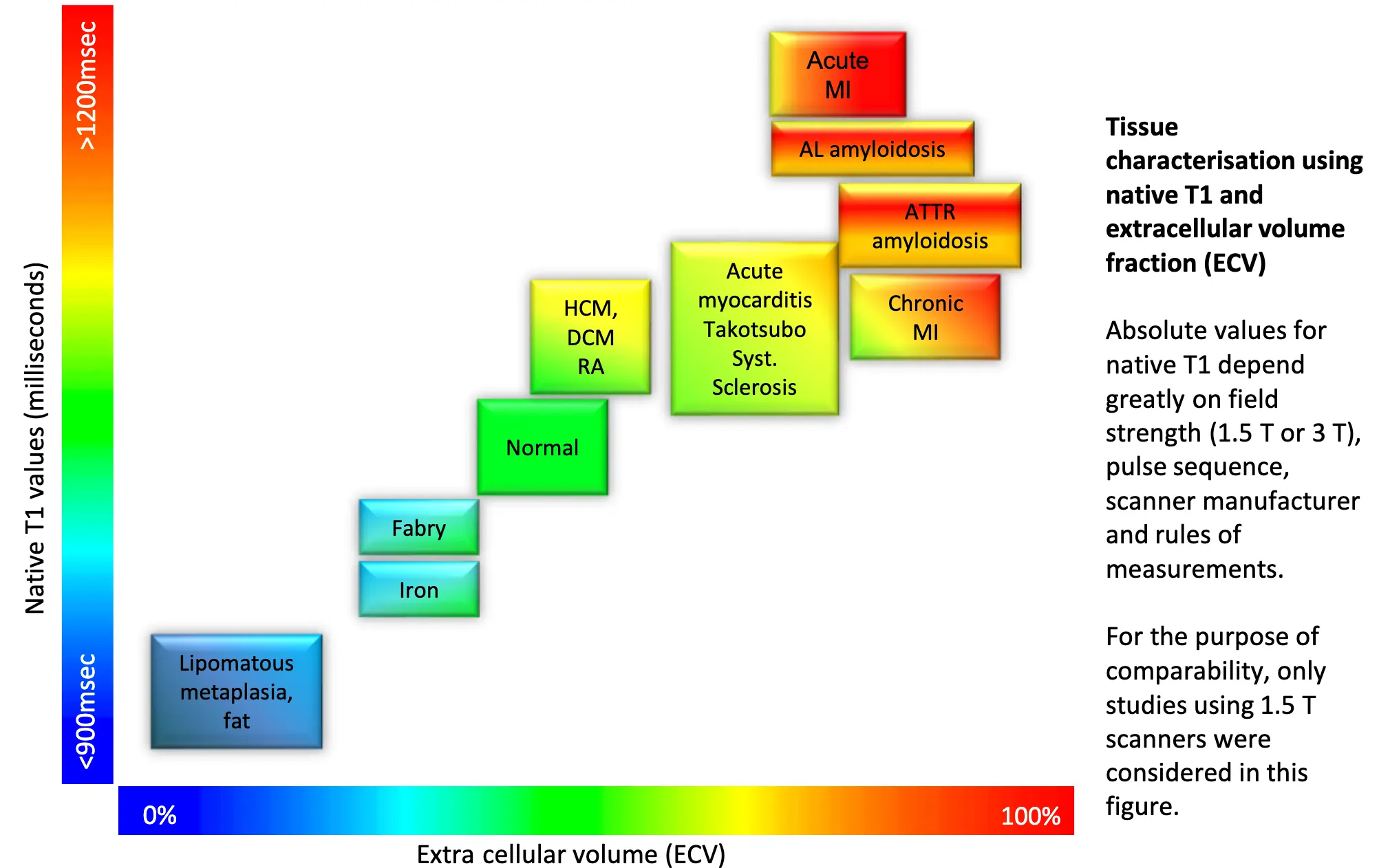 Figure source: page 42 of EACVI pocket guide
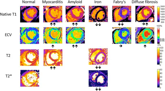
Typical appearance of T1, T2, T2*, and ECV maps in healthy subjects and in patients with myocardial disease. Arrows denote relative change in respective parametric maps.1
| T1 (native) | ECV | T2 | T2* | ||
|---|---|---|---|---|---|
| Infiltration | Iron overload | + | ? | + | ++ |
| Amyloid | ++ | ++ | ? | – | |
| Anderson-Fabry | ++ | – | + | – | |
| Acute myocardial injury | Edema | ++ | + | ++ | ? |
| Necrosis | ++ | ++ | + | ++ | |
| Hemorrhage | + | ? | + | ++ | |
| Fibrosis | Diffuse/global | + | ++ | ? | – |
| Focal/regional | + | ++ | – | – | |
| Clinical utility of parametric mapping techniques ordered by pathophysiologic mechanism and tissue characteristics. ++ = useful; + = potentially useful;? = unknown; − = not useful. Diffuse/global refers to findings affecting the majority of the myocardium, whereas focal/regional refers to localized, including patchy abnormalities. |
Footnotes
-
Messroghli DR, Moon JC, Ferreira VM, Grosse-Wortmann L, He T, Kellman P, Mascherbauer J, Nezafat R, Salerno M, Schelbert EB, Taylor AJ, Thompson R, Ugander M, van Heeswijk RB, Friedrich MG. Clinical recommendations for cardiovascular magnetic resonance mapping of T1, T2, T2* and extracellular volume: A consensus statement by the Society for Cardiovascular Magnetic Resonance (SCMR) endorsed by the European Association for Cardiovascular Imaging (EACVI). J Cardiovasc Magn Reson. 2017 Oct 9;19(1):75. doi: 10.1186/s12968-017-0389-8. Erratum in: J Cardiovasc Magn Reson. 2018 Feb 7;20(1):9. doi: 10.1186/s12968-017-0408-9. PMID: 28992817; PMCID: PMC5633041. ↩