- The aortic root is an extension of the LVOT, extending from the basal attachment of the aortic valve cusps within the LVOT to their peripheral attachment at the level of the sinotubular junction. Its components are the sinuses of Valsalva, the fibrous interleaflet triangles and the valvular cusps themselves.
The aortic root is a geometrically complex structure that includes:
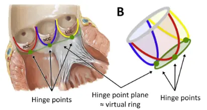
- the aortic valve annulus,
- not a true or distinct anatomic structure but is a virtual ring that may be defined by joining the basal attachments, or nadirs, of the three aortic leaflets. The distal (uppermost) attachments of the leaflets, in the shape of a crown, form a true anatomic ring.
- the interleaflet triangles,
- the semilunar aortic leaflets and their attachments,
- the aortic sinuses of Valsalva, and
- the sinotubular junction.
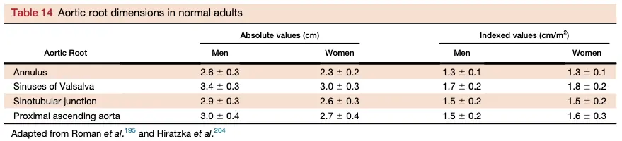
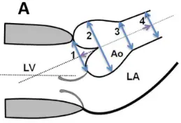
- Aortic measurements should be made at the following sites:
- the aortic valve annulus,
- the maximal diameter of the sinuses of Valsalva,
- the sinotubular junction (usually a demarcated transition between the sinuses of Valsalva and the tubular portion of the ascending aorta), and
- the maximal diameter of the proximal ascending aorta, including a notation of the distance between the measurement site and the sinotubular junction.
- When in the cardiac cycle should you make these aortic root measurements?
- mid-systole for aortic valve annulus
- measurements of the aortic annulus should be made in the zoom mode using standard electronic calipers in midsystole, when the annulus is slightly larger and rounder than in diastole, between the hinge points of the aortic valve leaflets (usually between the hinge point of the right coronary cusp and the edge of the sinus at the side of the commissures between the left coronary cusp and the noncoronary cusp) from inner edge to inner edge.
- end-diastole for all other aortic root measurements
- All other aortic measurements should be made at end-diastole, in a strictly perpendicular plane to that of the long axis of the aorta.
- mid-systole for aortic valve annulus
Aortic Valve Annulus
- The aortic annulus is defined as the luminal contour within a virtual plane aligned with the most basal attachment points of the three aortic valve cusps (sometimes referred to as the ‘basal hinge points’).
- Quantitative assessment requires the accurate identification of each of these three points in turn to create a plane that transects all three.
Sinuses of Valsalva
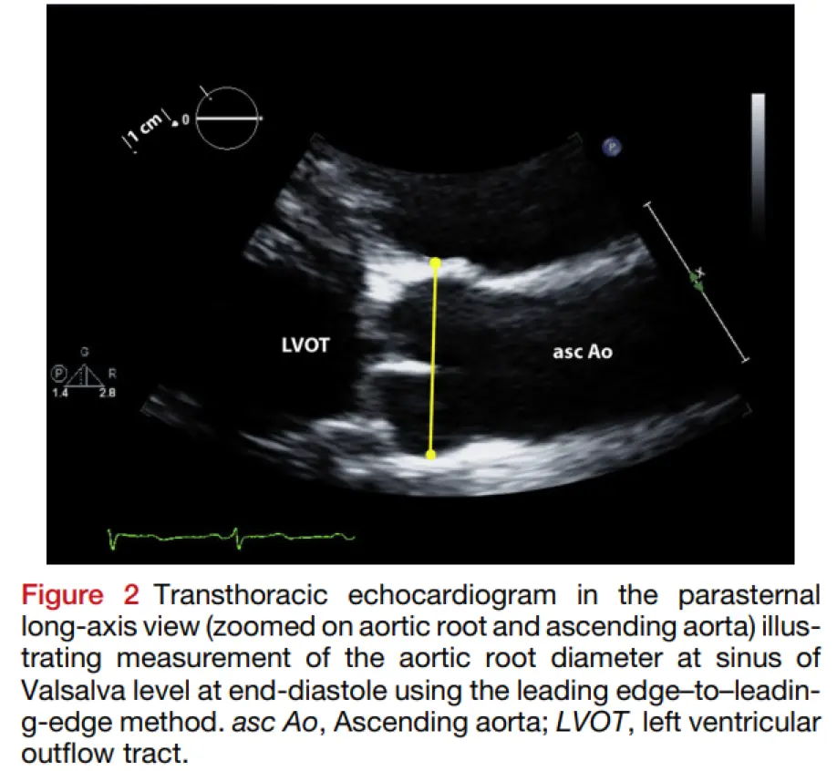
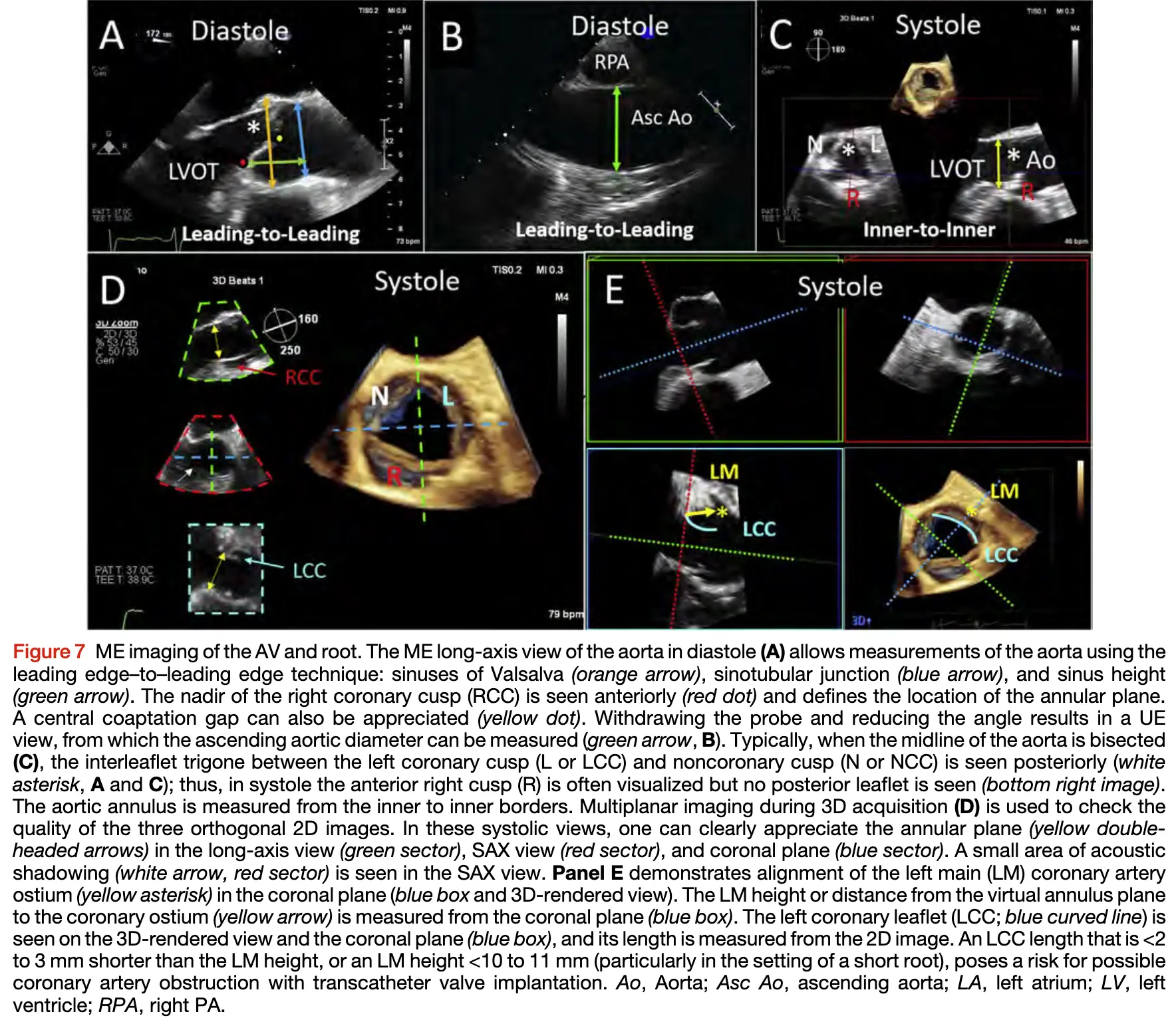
- The aortic root diameter at the sinus of Valsalva is measured in the PLAX view using the leading edge to leading edge method (from the leading edge of the anterior root wall to the leading edge of the posterior aortic root wall) and is measured at end-diastole (end-diastole can be recognized as being at the onset of the QRS).1
- Leading edge to leading edge method is kind of a relic of the past as it was initially devised to minimize the impact of ‘‘blooming’’ of bright reflectors on this measurement.1
- With TEE, aortic measurements are performed from the mid-esophageal (ME) long-axis view at end-diastole, using the leading edge-to-leading edge technique 2
Sinus of Valsalva Aneurysms
| ACCF/AHA Guidelines (2010) | ESC Guidelines (2014) | CCS Guidelines (2014) | |
|---|---|---|---|
| Degenerative | 5.5 cm | 5.5 cm | 5.5 cm |
| Bicuspid Aortic Valve | 5.5 cm | 5.0 cm | 5.0-5.5 |
| Marfan Syndrome | 4.0-5.0 cm | 5.0 cm | 5.0 cm |
| Other Genetic Syndromes† | 4.0-5.0 cm | — | 4.0-5.0 cm |
| Concomitant Cardiac Surgery | 4.5 cm | 4.5 cm | — |
| Annual Growth Rate | 0.5 cm/year | 0.3 cm/year | 0.5 cm/year |
 Figure and Table source
Reference Ranges