Doppler frequency changes depending on the direction and the velocity of a moving object, which in the case of cardiac echocardiography tends to involve RBCs in circulation or myocardial tissue (e.g. mitral annulus).
“When an ultrasound beam with known frequency () is reflected by the red blood cells (RBCs) or myocardial tissue, the frequency of the reflected ultrasound waves () increases when the red blood cells or the myocardium is moving toward the source of ultrasound. Conversely, the frequency of reflected ultrasound waves decreases when the red blood cells are moving away from the source. The change in frequency between the transmitted sound and the reflected sound is termed the frequency shift () or the Doppler shift (). The Doppler shift depends on the transmitted frequency (), the velocity of the moving target (), and the angle () between the ultrasound beam and the direction of the moving target as expressed in the Doppler equation.” (Oh, Chapter 4)
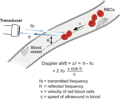 “If the angle θ is 0 degree (i.e., the ultrasound beam is parallel with the direction of blood flow), the maximal frequency shift is measured because the cosine of 0 degree is 1. Note that as angle θ increases, the corresponding cosine becomes progressively less than 1, and this will result in underestimation of the Doppler shift (Δf) and hence peak velocity.” (Oh, Chapter 4)
Doppler Pattern Recognition
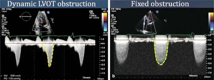
Obstruction
- Dynamic LVOT obstruction
- Fixed obstruction
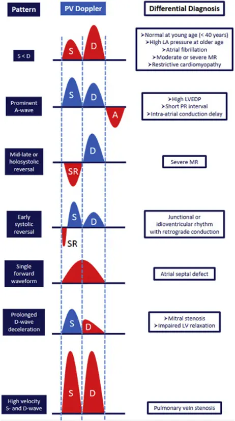
Pulmonary Vein Doppler
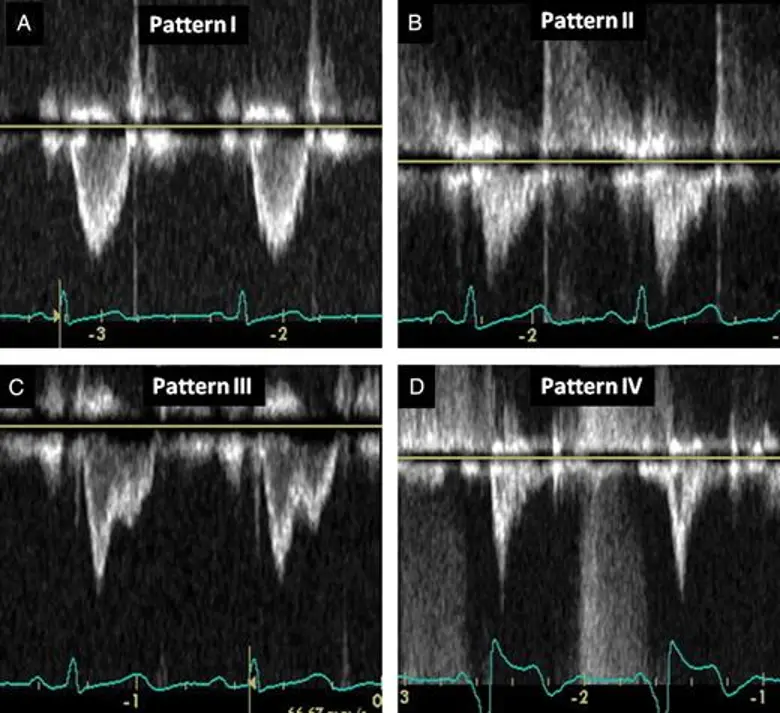
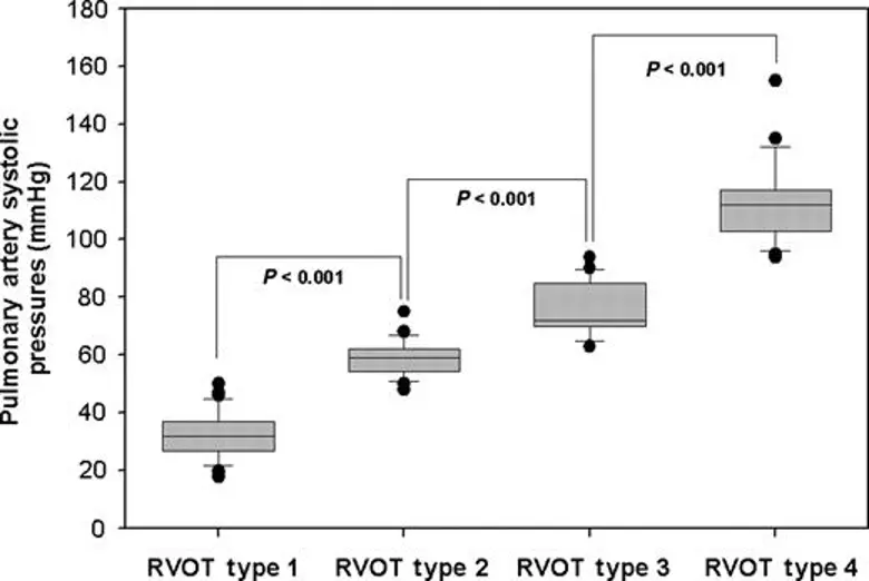
RVOT VTI Doppler Patterns
- “Visual assessment of RVOT spectral signals demonstrates the presence of four dynamic patterns, independent of the etiology of the Pulmonary Hypertension, that not only correlate with the severity of Pulmonary Hypertension, but also are useful in identifying a range of PASP with great accuracy that minimizes subjective interpretation.” 1
Footnotes
-
Angel López-Candales, Kathy Edelman, Shape of the right ventricular outflow Doppler envelope and severity of pulmonary hypertension, European Heart Journal - Cardiovascular Imaging, Volume 13, Issue 4, April 2012, Pages 309–316, https://doi.org/10.1093/ejechocard/jer235 ↩