To interpret a nuclear study there are multiple programs and steps that need to be followed to ensure accurate interpretation and complete dictation. When logging in for the day I will open all four programs located below to setup my workstation. Below is an outline
Cedars Terminal (CSI)
- Located on all computers in nuclear lab as well as the CT reading swing office in the hallway
- Login information:
- Username: CVfellow
- Password: (ask on the fellows WhatsApp)
- Locate study by searching patient name in the top
- Once patient is selected, bottom of screen will populated the associated nuclear study files
- Locate the QP/QS file and left click to select it
- After selecting/highlighting the above file, in the top left find and click the red QP/QS icon to open the study
- It is important to open all studies through this process to ensure the processing from the tech is correctly applied to your study. Otherwise the images you review may look different compared to what your staff will open when you present your study!
CardioServ
- All studies have an associated EKG strip that needs to be interpreted into the first box of your study interpretation
- There are multiple ways to access the cardio server
- Desktop icon for Cardioserv – should be on most computers in the nuclear lab
- Epic client → Clinical references (CTL +8) → Cardioserv
- SLH Access Citrix Hub → Apps, all → CardioServ
- 📝 not all fellows will have this app in Citrix. You can request access using eRequest.
- Open CardioServ – by whichever preferred method – p# and password will auto populate
- Setup the inbox in the top left with the following parameters
Site: AllType: STRESSStatus: Unconfirmed- Search by: Name
- Then search the patient name in the below box and open the associated strips
- 💡Tip: You can use the page down button and the right arrow to navigate through the especially long strips to increase speed.

Syngo Via
- Low resolution CT for attenuation correction and Cardioscan CTs are reviewed through syngo.via
- Locate “syngo.via Single Sign On” on the Desktop – open this link
- Search for your patient by last name and double click the study to open
Dragon Dictation
- Open dragon by way of the “Dragon Medical One” icon on the desktop or via epic client
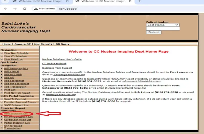
Nuclear Database
- The Chrome desktop shortcut labeled “Nuclear Database” is how you navigate to your study dictation
- Open the shortcut – your P# and password will automatically populate and log you in.
- All current fellows have been granted access. Please email Rob if there is any issues with logging in.
- Navigate to the
Phys Dictation Liston the left hand side (towards the bottom) - Clicking the list will open you to the reading list for prepared/ pending studies of the day separated out by types:
PET,Stress first SPECTand2 Day SPECT - Search for your patient name in the list and click to open the dictation for your study
- Utilize the Dragon Dictation to populate a template for your study
- Click
Savein the bottom left after completing your dictation - All of the fellow pended studies will now be found at the bottom of the “Phys Dictation List”.
- When staff is reviewing, they will open the study, make edits and click the drop down box to change from incomplete to complete which will push the study to Epic.
Reporting Templates
The following examples are taken (and slightly modified) from 1
Pharmacologic MPI
*** (Single/2 day) Rest/Stress *** (or Stress/Rest) Pharmacologic Stress Myocardial Perfusion Imaging with LV function analysis

Indication

(select one) (Diagnosis of coronary artery disease/known coronary artery disease/chest pain/shortness of breath/Preoperative assessment/Evaluation of myocardial viability/Risk Stratification/Other)

Clinical history

X-year-old man/women with a history of:

Cardiac History:

Cardiac Risk Factors:

Prior cardiac imaging and procedures:

Current symptoms:

Technique

At rest, the patient received x mCi of x tracer. X minutes later, resting tomographic images of the heart were obtained.

Pharmacologic stress testing was performed with adenosine/dipyridamole/dobutamine/regadenoson at a rate of ____ for ___minutes. Additionally, low-level exercise was performed along with the vasodilator infusion (specify: ____). Resting HR was x with a peak heart rate of x bpm and x% maximum predicted heart rate . The rest blood pressure was ___ mm/Hg and increased/decreased to ___ mm/Hg, which is a normal/hypotensive/hypertensive response. The patient developed significant symptoms, which included ____.

The resting EKG showed x with no significant ST/T abnormalities that would preclude interpretation. The stress EKG showed (no) ST-segment changes consistent with myocardial ischemia, with x mm horizontal/upsloping/downsloping ST depression in the x leads. ST depressions began at x min of rest/stress and resolved at x min of rest/stress.

At peak stress, the patient received x mCi of x. Stress tomographic imaging was performed x minutes later. The rest and post-stress images were acquired with ECG gating, for assessment of left ventricular systolic function. All imaging was performed on a x camera and data were analyzed using x software.

Findings

The overall quality of the study is poor/fair/good/excellent. Review of the raw imaging demonstrates (no) significant motion during stress/rest image acquisition. Attenuation artifact was present/absent in the x walls.

Review of the perfusion images shows symmetric or improved uptake of tracer in all portion of the left ventricle from rest to stress imaging OR show an x severity x sized perfusion defect in the anterior wall that is x reversible, a x sized x severity perfusion defect in the lateral wall that is x reversible, and a x sized x severity perfusion defect in the inferior wall that is x reversible. Quantitative evaluation shows a summed stress score of x, a summed rest score of x, and a summed difference score of x. This represents a myocardial ischemic fraction of x%.

Gated SPECT images shows that the left ventricle is normal/enlarged in size and shows normal systolic performance. The LVEF at rest is x% and x% on post-stress images. No regional wall motion abnormalities are present during either stress or rest imaging.

Transient ischemic dilation, a high-risk marker, is/is not present. Left ventricular/right ventricular hypertrophy is/is not present. Left ventricular/right ventricular dilation is/is not present.

Impression

1. Myocardial perfusion imaging is normal with no evidence of ischemia or scar OR Myocardial perfusion imaging is abnormal with a small/moderate/large area of ischemia/infarction in the distribution of the x artery.
2. Left ventricular systolic function is normal/abnormal with (no)/x regional wall motion abnormalities. Left/Right ventricular hypertrophy/dilation is present.
3. In comparison with the previous study of x date, there has been (no)/a change in left ventricular perfusion, size, or function.
Exercise MPI
*** (Single/2 day) Rest/Stress *** (or Stress/Rest) Exercise Stress Myocardial Perfusion Imaging with LV function analysis

Indication
*** (select one) (Diagnosis of coronary artery disease/known coronary artery disease/chest pain/shortness of breath/Preoperative assessment/Evaluation of myocardial viability/Risk Stratification/Other)

Clinical history:
X-year-old man/women with a history of ***

Cardiac History: ***

Cardiac Risk Factors: ***

Prior cardiac imaging and procedures: ***

Prior nuclear stress test date: ***

Current symptoms: ***

Technique

At rest, the patient received *** mCi of *** tracer. *** minutes later, resting tomographic images of the heart were obtained.

The patient then underwent *** exercise treadmill/bike stress testing according to the *** protocol, exercising for *** minutes, achieving a workload of *** metabolic equivalents (METS). Resting HR was *** with a peak heart rate of *** bpm and ***% maximum predicted heart rate and rate-pressure product of ***. Resting BP was ***/*** mm Hg and Peak BP was ***/*** mm Hg, which is a *** normal/hypertensive/hypotensive response. The heart rate response to recovery was *** normal/abnormal. *** The test was terminated due to chest pain/shortness of breath/fatigue/leg pain. Other symptoms included ***.

The resting EKG showed *** with no significant ST/T abnormalities that would preclude interpretation. The stress EKG showed *** (no) ST-segment changes consistent with myocardial ischemia, *** with *** mm horizontal/upsloping/downsloping ST depression in the *** leads. ST depressions began at *** min of rest/stress and resolved at *** min of rest/stress. The Duke Treadmill score was ***, predicting a *** low/intermediate/high risk.

At peak stress, the patient received *** mCi of ***. Stress tomographic imaging was performed *** minutes later. The rest and post-stress images were acquired with ECG gating, for assessment of left ventricular systolic function. All imaging was performed on a *** camera and data were analyzed using *** software.

Findings

The overall quality of the study is *** poor/fair/good/excellent. Review of the raw imaging demonstrates *** (no) significant motion during stress/rest image acquisition. Attenuation artifact was *** present/absent in the *** walls.

Review of the perfusion images shows symmetric or improved uptake of tracer in all portions of the left ventricle from rest to stress imaging *** shows an *** severity, ***-sized perfusion defect in the *** (anterior/lateral/inferior) wall that is *** reversible. Quantitative evaluation shows a summed stress score of ***, a summed rest score of ***, and a summed difference score of ***. This represents a myocardial ischemic fraction of ***%.

Gated *** (SPECT/PET) images show that the left ventricle is *** normal/enlarged in size and shows normal systolic performance. The LVEF at rest is ***% and ***% on post-stress images. *** No regional wall motion abnormalities are present during either stress or rest imaging.

Transient ischemic dilation, a high-risk marker, *** is/is not present. *** Left ventricular/right ventricular hypertrophy *** is/is not present. *** Left ventricular/right ventricular dilation *** is/is not present.

Impression

1. *** (Myocardial perfusion imaging is normal with no evidence of ischemia or scar/Myocardial perfusion imaging is abnormal with a *** small/moderate/large area of *** ischemia/infarction in the distribution of the *** artery).
2. Left ventricular systolic function is *** (normal/abnormal) with *** (no/***) regional wall motion abnormalities. *** Left/Right ventricular *** hypertrophy/dilation is present.
3. In comparison with the previous study of *** date, there has been *** (no/a) change in left ventricular perfusion, size, or function.
Exercise MPI with Combined Conclusion
Reason for Study: Preoperative evaluation prior to non-cardiac surgery.

Clinical History: Mr. [XXXXXX] is a 56-year-old male with a history of hypertension and dyslipidemia with no prior known coronary artery disease who is currently asymptomatic. He has not had prior coronary angiography and has a SPECT myocardial perfusion imaging study from [xx/xx/xxxx] for comparison.

Stress ECG: (not provided in this appendix for brevity).

Isotope Administration

This was a gated SPECT myocardial perfusion imaging study. A one-day rest-stress imaging protocol was followed. The isotope used for imaging was 99mTc-sestamibi. Rest imaging was performed after an injection of 7.1 mCi. Stress imaging was performed after an injection of 21.3 mCi.

Nuclear Stress Findings

Nuclear Study Quality

Overall imaging quality was good.

Perfusion Conclusion

LV perfusion is probably normal.

Perfusion Defect #1

There is a small region with moderate reduction in uptake in the apical to mid anterior segment(s) that is predominately reversible. There is normal wall motion in the defect area. The defect appears to be shifting breast artifact, but ischemia cannot be ruled out. The perfusion defect is visually present but not quantitatively significant.

Perfusion Comments

There is no evidence of transient ischemia dilation (TID). The rest study indicates well-preserved viability.

Function Comments

Left ventricular function post-stress was normal with an ejection fraction of 63%. The stress end-diastolic cavity size was normal (52 mL/m2). The stress end-systolic cavity size was normal (19 mL/m2).

Interpretation Summary

- The stress electrocardiogram was positive for electrocardiographic evidence of myocardial ischemia.
- The Duke Treadmill Score was intermediate risk at -5.
- The patient developed typical angina at peak stress.
- LV perfusion is probably normal.
- The small region with moderate reduction in uptake in the apical to mid anterior segment(s) appears to be shifting breast artifact but ischemia cannot be ruled out.
- Left ventricular function post-stress was normal with an ejection fraction of 63%.

Nuclear and Stress Combined Conclusion

The ECG and SPECT portions of the stress study are discordant, but the following factors support an intermediate risk of inducible myocardial ischemia:

- Poor exercise workload achieved during stress.
- Anginal symptoms during stress.
- Multiple cardiovascular risk factors.

Further cardiac evaluation for ischemic heart disease could be considered, especially in the setting of progressive or typical angina.

Nuclear Prior Study

Compared with the prior study dated [xx/xx/xxxx], the perfusion defect is new. There has been no significant change in left ventricular function.
Footnotes
-
Tilkemeier PL, Bourque J, Doukky R, Sanghani R, Weinberg RL. ASNC imaging guidelines for nuclear cardiology procedures. Journal of Nuclear Cardiology. 2017;24(6):2064-2128. doi:10.1007/s12350-017-1057-y ↩
