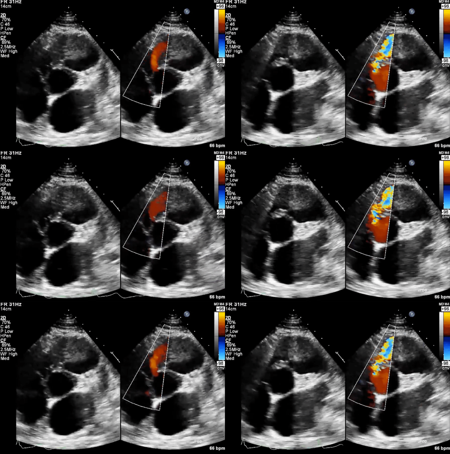
- Dilates the Left side of the heart
- to dilate a chamber, you need to fill it during diastole
- systole: LV contracts → blood travels through VSD into RV and into the pulmonary arteries.
- Due to flow dynamics of the VSD, the AV and/or TV can be “sucked into” the VSD → aortic regurgitation and/or tricuspid regurgitation
- ECG
- LAE or LVH (if hemodynamically significant VSD)
- 4 types of VSD by location
- membranous VSD (perimembranous)
- inlet VSD (atrioventricular canal defect)
- often associated with primum ASD and cleft MV
- muscular VSD
- subpulmonary VSD (subarterial)
- found just beneath the pulmonary valve
- most common in patients of Japanese 🇯🇵 descent
-1754868393010.webp)
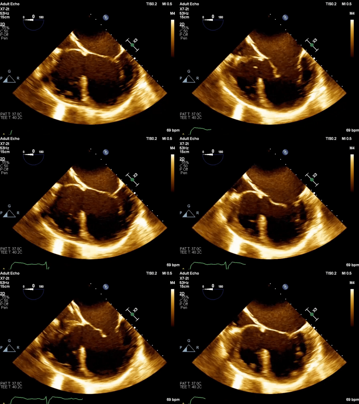
Echo in VSD
-1754871370645.webp)
-
Membranous VSD
-
Inlet VSD
Management
- Sources:
Muscular VSD
-1754869604185.webp)
Non-Muscular VSD
-1754869635605.webp)
Unrepaired VSD in Adult
-1754870091671.webp)
Repaired VSD in Adult
-1754870123729.webp)

