Echo of LAA
- On TEE, imaging of the LAA is recommended to be obtained at ME 0˚, 45˚, 90˚, and 135˚; add X-plane views PRN
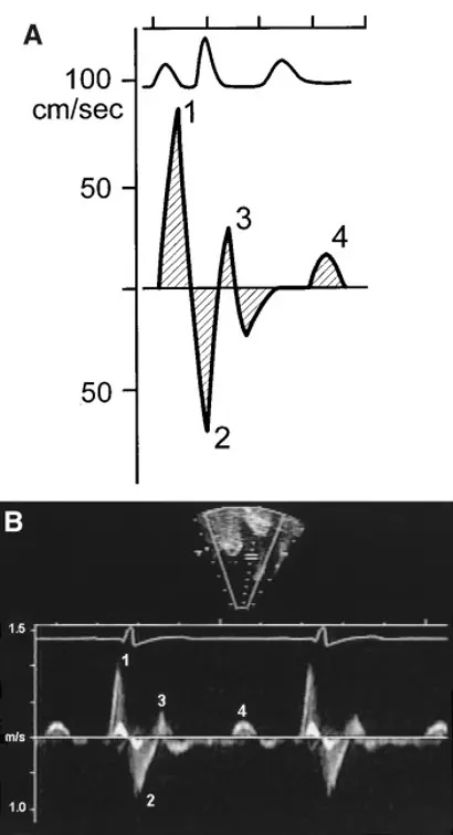
Emptying Velocities on TEE
CT of LAA
- “Cardiac computed tomography, particularly when delayed imaging is performed, is a reliable alternative to TEE for the detection of LA/LAA thrombi/clot, avoiding the discomfort and risks associated with TEE.” 1
- “In a subanalysis of studies in which delayed imaging was performed, the diagnostic accuracy significantly improved to a mean weighted sensitivity and specificity of 100% and 99%, respectively, whereas the positive predictive value and negative predictive value increased to 92% and 100%, respectively. The accuracy for this technique was 99%.”
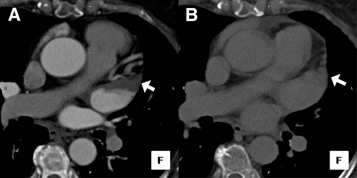 Figure source: 2 - Axial image of a Cardiac CT Study Performed in a Patient Before RF Pulmonary Vein Ablation: (A) Initial image obtained after contrast injection demonstrates filling defect in the left atrial appendage tip. (B) Delayed image obtained 1 min after initial scan shows homogeneous opacification, suggesting an absence of thrombus.
Footnotes
-
Romero J, Husain SA, Kelesidis I, Sanz J, Medina HM, Garcia MJ. Detection of left atrial appendage thrombus by cardiac computed tomography in patients with atrial fibrillation: a meta-analysis. Circ Cardiovasc Imaging. 2013 Mar 1;6(2):185-94. doi: 10.1161/CIRCIMAGING.112.000153. Epub 2013 Feb 13. PMID: 23406625. ↩
-
Garcia MJ. Detection of Left Atrial Appendage Thrombus by Cardiac Computed Tomography. JACC: Cardiovascular Imaging. 2009;2(1):77-79. doi:10.1016/j.jcmg.2008.10.003 ↩